
Learn names through pictures
Cell organelles name typing
Look at the pictures and type the names of cell organelles.
Explore 16 names, one at a time.
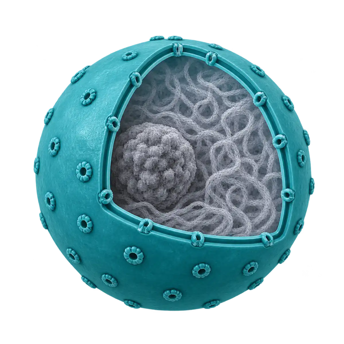
cell organelles typing guide →
About Cell organelles name typing
Read names from Nucleus and Nucleolus to storage-related structures using representative cell models. This set includes both membrane-bound organelles and major non-membranous structures such as Ribosome and Centriole. Every name is visible, so reading microscopy images is unnecessary. Look at internal details and gray transport context as you copy the biological vocabulary one structure at a time.
The 16 entries include Rough endoplasmic reticulum, Smooth endoplasmic reticulum, Golgi apparatus, Ribosome, Transport vesicle, Endosome, Lysosome and Peroxisome. Mitochondrion, Centrosome and Centriole lead to Chloroplast, Central vacuole and Amyloplast. Animal-cell, plant-cell and plant-storage models share a vocabulary list, not a claim that all 16 must occur together in one cell.
Surface ribosomes distinguish rough from smooth endoplasmic reticulum in the models. A centrosome with its centriole pair remains different from a single centriole. Mitochondrial cristae and chloroplast grana are not interchangeable details. Transport vesicles, endosomes, lysosomes and peroxisomes cannot be conclusively identified in real material by a simple outline or color, so read the depicted contents and trafficking context as well as the complete label.
This learning draft awaits specialist cell-biology review. Images are conceptual illustrations rather than micrographs, with no common scale or natural-color claim. A crystalline peroxisomal core represents only some cells. Amyloplast denotes a starch-storage plastid rather than a chloroplast. The collection is not a complete map of one cell and provides no definitive identification rule for real organelles.
The first nonempty input for Nucleus starts the timer. When Nucleolus or another new label appears, read its picture and complete name before continuing. A matching answer advances without Enter, and unfinished text can be edited in place. Enter on an incorrect nonempty answer adds one error and clears that attempt. After Amyloplast, review time, keystrokes per minute and errors; the best time stays in this browser by topic, set and language.
How to play
- Open the basic 16 cell organelles names and read the first label, Nucleus, with its selected area.
- Copy a complete label such as Nucleolus, or an alternative registered for that particular entry.
- A matched cell organelles name advances automatically; pause to read the new illustration before continuing.
- Edit an unfinished Nucleus answer directly. Enter on incorrect nonempty text counts one error and clears the field.
- Finish at Amyloplast, review the statistics and retry the same 16-name sequence as a new round.
Who is it for?
- Readers beginning cell organelles vocabulary who want a visible name such as Nucleus beside a picture.
- Learners comparing the complete spelling and scope of longer labels such as Nucleolus.
- Classes or study partners using a fixed sequence of 16 cell organelles names for a short typing activity.
- People who prefer to reach Amyloplast at their own pace and compare personal time and errors on a retry.
Practice sets
- Basic 16 · 16 names
Copy 16 representative names in order from Nucleus to Amyloplast, comparing the spelling and selected range of terms such as Nucleolus.
Names and spelling
Surface ribosomes distinguish rough from smooth endoplasmic reticulum in the models. A centrosome with its centriole pair remains different from a single centriole. Mitochondrial cristae and chloroplast grana are not interchangeable details. Transport vesicles, endosomes, lysosomes and peroxisomes cannot be conclusively identified in real material by a simple outline or color, so read the depicted contents and trafficking context as well as the complete label.
The actual display list is Nucleus · Nucleolus · Rough endoplasmic reticulum · Smooth endoplasmic reticulum · Golgi apparatus · Ribosome · Transport vesicle · Endosome · Lysosome · Peroxisome · Mitochondrion · Centrosome · Centriole · Chloroplast · Central vacuole · Amyloplast. Each label is an independent entry; a containing structure does not automatically answer for its parts. Use the current-language display name or a specifically registered alternative. The game does not infer arbitrary synonyms, abbreviations or translations. Letter case, spaces and supported punctuation are normalized; Latin accent marks can be omitted and ß can be entered as ss, without changing meaningful word order or numbers.
This learning draft awaits specialist cell-biology review. Images are conceptual illustrations rather than micrographs, with no common scale or natural-color claim.
Practice tips
- Surface ribosomes distinguish rough from smooth endoplasmic reticulum in the models.
- A centrosome with its centriole pair remains different from a single centriole.
- Mitochondrial cristae and chloroplast grana are not interchangeable details.
- Transport vesicles, endosomes, lysosomes and peroxisomes cannot be conclusively identified in real material by a simple outline or color, so read the depicted contents and trafficking context as well as the complete label.
- Copy a complete label such as Nucleolus, or an alternative registered for that particular entry.
- Finish at Amyloplast, review the statistics and retry the same 16-name sequence as a new round.
Frequently asked questions
Which cell organelles names can I practice?
Read names from Nucleus and Nucleolus to storage-related structures using representative cell models. This set includes both membrane-bound organelles and major non-membranous structures such as Ribosome and Centriole.
What does the list from Nucleus to Amyloplast cover?
The 16 entries include Rough endoplasmic reticulum, Smooth endoplasmic reticulum, Golgi apparatus, Ribosome, Transport vesicle, Endosome, Lysosome and Peroxisome. Mitochondrion, Centrosome and Centriole lead to Chloroplast, Central vacuole and Amyloplast.
What distinctions matter when reading Nucleolus and related names?
Surface ribosomes distinguish rough from smooth endoplasmic reticulum in the models. A centrosome with its centriole pair remains different from a single centriole. Mitochondrial cristae and chloroplast grana are not interchangeable details.
What are the limits of the cell organelles illustrations?
Amyloplast denotes a starch-storage plastid rather than a chloroplast. The collection is not a complete map of one cell and provides no definitive identification rule for real organelles.
Which written answers work in the cell organelles set?
Use the current-language display name or a specifically registered alternative. The game does not infer arbitrary synonyms, abbreviations or translations. Letter case, spaces and supported punctuation are normalized; Latin accent marks can be omitted and ß can be entered as ss, without changing meaningful word order or numbers.
Where is my result after Amyloplast stored?
Enter on an incorrect nonempty answer adds one error and clears that attempt. After Amyloplast, review time, keystrokes per minute and errors; the best time stays in this browser by topic, set and language.
This learning draft awaits specialist cell-biology review. Images are conceptual illustrations rather than micrographs, with no common scale or natural-color claim. A crystalline peroxisomal core represents only some cells. Amyloplast denotes a starch-storage plastid rather than a chloroplast. The collection is not a complete map of one cell and provides no definitive identification rule for real organelles.